Veterinary Imaging for Foreign Body Detection
Foreign body ingestion is a common issue in veterinary medicine that can lead to serious complications if not detected and treated promptly. Veterinary imaging techniques provide critical diagnostics by visualizing the location, size, shape, and composition of foreign bodies within an animal's gastrointestinal tract or other organs.
The primary tools for detecting foreign bodies include radiography (X-rays), ultrasound, computed tomography (CT) scans, and magnetic resonance imaging (MRI). Each method has its strengths:
- Radiography: Provides basic images of metallic objects or dense materials. It is quick but may miss softer foreign bodies.
- Ultrasound: Useful for assessing soft tissues and detecting less dense materials such as bones, stones, or certain plastics. It can also provide real-time imaging during procedures like endoscopy.
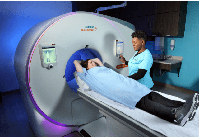
- CT Scans: Offer higher resolution than standard radiographs, making them ideal for identifying complex shapes and compositions of foreign bodies. They are particularly useful in cases involving metal objects or when a detailed cross-sectional view is necessary.
- MRI: Although less commonly used for detecting foreign bodies due to its lower contrast with metallic materials, MRI can be valuable for assessing soft tissue damage surrounding the foreign body.
When conducting these imaging procedures, it is crucial to prepare the animal properly. This may involve fasting the patient, administering mild sedation or anesthesia if necessary, and ensuring the animal lies still during the scan to obtain clear images. The imaging process itself requires precise calibration of equipment and careful interpretation by experienced radiologists.
The data obtained from these tests is essential for determining the appropriate course of treatment. Depending on the findings, veterinarians may recommend surgical removal, endoscopic retrieval, or other interventions based on the type and location of the foreign body. Early detection can prevent severe complications such as perforation, infection, and obstruction.
Given the critical nature of these tests, it is important to work with laboratories that have a proven track record in diagnostic imaging. These labs should adhere strictly to international standards (ISO 15408 for radiography, IEC 62304 for software used in medical devices) and maintain accreditation from reputable bodies such as the American College of Veterinary Radiology or the Royal College of Veterinary Surgeons.
Scope and Methodology
| Procedure | Description |
|---|---|
| Radiography (X-ray) | Captures images of metallic and dense objects. Suitable for quick initial screening. |
| Ultrasound | Provides real-time imaging, particularly useful for soft tissues and less dense materials. |
| CT Scans | Offers high-resolution images of complex shapes and compositions. Ideal for detailed analysis. |
| MRI | Useful for soft tissue damage assessment, especially in cases involving non-metallic materials. |
Customer Impact and Satisfaction
- Quick Diagnosis: Timely detection helps prevent life-threatening complications.
- Pain-Free Procedure: Sedation ensures the animal remains comfortable throughout the process.
- High Accuracy: Advanced equipment and skilled operators ensure reliable results.
Environmental and Sustainability Contributions
- Eco-Friendly Equipment: Our imaging systems comply with environmental standards to minimize waste and energy consumption.
- Reduced Animal Stress: Sedation techniques reduce the stress on animals, promoting a more humane approach to diagnosis.


