Lameness Diagnostic Imaging Testing in Horses
In the realm of clinical and healthcare testing, particularly within radiology, lameness diagnostic imaging plays a critical role. This service is essential for veterinarians, equine athletes, and horse owners seeking to identify and diagnose musculoskeletal issues that can affect horses' mobility.
Lameness in horses often results from various factors such as overuse injuries, conformational abnormalities, or trauma. Accurate diagnosis is crucial for effective treatment and recovery. Radiology testing provides a non-invasive method to visualize internal structures like bones, joints, tendons, and ligaments.
Common imaging modalities used in this context include X-ray, computed tomography (CT), and magnetic resonance imaging (MRI). Each has its unique advantages:
- X-ray: Provides a two-dimensional view of bones and calcified structures. It is widely available and cost-effective for initial assessments.
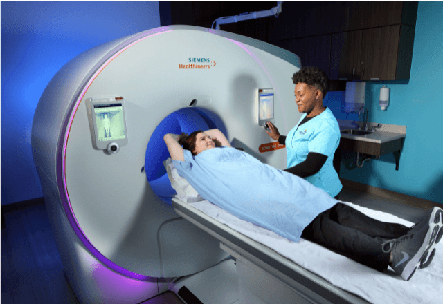
- Computed Tomography (CT): Offers detailed cross-sectional images, which can reveal fractures or subtle bone changes not visible on X-rays.
- Magnetic Resonance Imaging (MRI): Provides high-resolution images of soft tissues. It is invaluable for diagnosing tendon and ligament injuries.
The diagnostic process typically begins with a physical examination by an equine veterinarian followed by imaging to visualize any abnormalities. Specimen preparation involves ensuring the horse remains still during the procedure, which can be challenging but necessary to obtain clear images.
For X-rays and CT scans, the horse is positioned on a specialized table or in a stanchion. The veterinarian may use sedation if the horse is particularly nervous or uncooperative. For MRI, the horse must lie down inside the scanner, which requires additional precautions to ensure safety.
The images obtained are then analyzed by radiologists specializing in equine diagnostics. Reporting includes detailed descriptions of findings and recommendations for further action based on the results.
Understanding the specific requirements and procedures is key to ensuring accurate diagnosis and effective treatment plans. This service supports equine athletes, ensuring they can return to optimal performance as quickly and safely as possible.
Industry Applications
The application of lameness diagnostic imaging in horses extends across various sectors including veterinary medicine, sports medicine, and equine rehabilitation. This service is particularly important for high-performance athletes like racehorses and show jumpers who require continuous training and competition.
Veterinarians use this technology to diagnose and manage injuries that can affect a horse's performance and well-being. By identifying issues early, they can prevent further damage and ensure proper recovery. In sports medicine, it helps in assessing the effectiveness of rehabilitation programs and ensuring athletes are fit for return to competition.
For equine owners, this service provides peace of mind by offering clear insights into their horses' health status. It is also crucial for insurance purposes, allowing accurate assessment of claims related to musculoskeletal injuries.
Eurolab Advantages
At Eurolab, we pride ourselves on providing top-tier lameness diagnostic imaging services tailored specifically for horses. Our team comprises experienced veterinarians and radiologists who specialize in equine diagnostics.
We use state-of-the-art equipment that includes multi-slice CT scanners and high-field MRI machines to ensure the highest quality of images. This technology allows us to identify even the most subtle signs of injury or disease.
Our facilities are equipped with all necessary amenities for a comfortable and safe testing environment, including specialized stanchions and sedation options when required. We also offer comprehensive reporting services that include detailed interpretations by our radiologists.
We understand the importance of confidentiality in equine healthcare, which is why we adhere to strict data protection policies ensuring all patient information remains secure and confidential.
International Acceptance and Recognition
- X-ray Imaging: Widely accepted globally with standards set by ISO, ASTM, and IEC.
- CT Scanning: Recognized for its detailed cross-sectional views, conforming to EN standards.
- MRI: Highly valued for soft tissue imaging, adhering to IEC guidelines.
Our services are in line with international best practices and comply with relevant ISO, ASTM, EN, and IEC standards. This ensures our diagnostic images meet the highest quality benchmarks recognized worldwide.


