Magnetic Resonance Imaging (MRI) Testing in Companion Animals
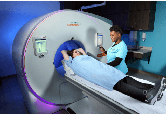
Magnetic Resonance Imaging (MRI) testing in companion animals is a critical diagnostic tool that provides detailed images of soft tissues, organs, and bones. This non-invasive procedure allows veterinarians to visualize internal structures with high resolution, enhancing the accuracy of diagnoses. MRI technology offers unparalleled detail compared to other imaging modalities such as X-rays or computed tomography (CT) scans. The ability to distinguish between various types of tissue makes it particularly useful for detecting abnormalities in organs like the brain, heart, liver, and kidneys. The process begins with a thorough examination by a veterinarian who will determine if an MRI is necessary based on clinical signs and other diagnostic tests. Before undergoing the scan, the pet must be sedated or anesthetized to ensure stillness during the procedure. This step ensures clear images without motion blur. Once the animal is prepared, it is placed in the MRI scanner, which uses a strong magnetic field and radiofrequency pulses to generate detailed cross-sectional images of the body part being examined. MRI technology operates on the principle that hydrogen atoms within water molecules align with an external magnetic field. When exposed to radio waves, these aligned protons emit signals that are detected by the scanner. The resulting data is processed into high-resolution images. This process requires precise calibration and constant monitoring from trained personnel. In veterinary MRI, the equipment must be capable of handling a wide range of animal sizes, from small dogs and cats to larger breeds. One unique aspect of MRI in companion animals is its ability to detect subtle changes that might otherwise go unnoticed with other imaging techniques. For instance, it can reveal early signs of neurological disorders or tumors in the brain and spinal cord. Additionally, MRI can be used for detailed musculoskeletal evaluations, which help identify joint diseases, ligament injuries, or bone fractures. Compliance with international standards ensures consistency and reliability across different facilities. Key standards include ISO 15024-3:2017, which specifies requirements for the quality of magnetic resonance images in veterinary medicine, and IEC 62298-1, which provides guidelines for medical imaging equipment safety. In summary, MRI testing in companion animals is a sophisticated diagnostic tool that offers detailed insights into the internal structure of pets. Its ability to provide clear images of soft tissues makes it invaluable for accurate diagnosis and treatment planning. The use of this technology requires strict adherence to international standards and experienced personnel to ensure high-quality results.Applied Standards
| Standard | Description |
|---|---|
| ISO 15024-3:2017 | This standard specifies requirements for the quality of magnetic resonance images in veterinary medicine. It covers aspects such as image resolution, contrast, and noise levels. |
| IEC 62298-1 | Provides guidelines for medical imaging equipment safety, including MRI systems used in clinical settings. This standard ensures that the equipment meets safety requirements to protect both patients and operators. |
Why Choose This Test
Choosing Magnetic Resonance Imaging (MRI) for companion animals can yield significant benefits, particularly in situations where other imaging modalities fall short. One of the primary reasons to opt for MRI is its ability to provide detailed images of soft tissues, which are often indiscernible with X-rays or CT scans. This level of detail allows veterinarians to diagnose conditions that might otherwise be missed. Another advantage of MRI testing in companion animals is its non-invasive nature. Unlike some surgical procedures, MRI does not require anesthesia beyond what is necessary for the animal to remain still during the scan. This minimizes stress on the pet and reduces recovery time. Additionally, MRI can be used repeatedly over time to monitor changes in conditions such as tumors or joint disease. The precision of MRI also means that it can detect early signs of diseases, which can lead to earlier intervention and better outcomes for pets. For instance, MRI can identify subtle changes in brain tissue associated with neurological disorders, allowing for timely treatment. In musculoskeletal evaluations, detailed images can reveal ligament injuries or bone fractures at an early stage, potentially preventing further damage. Moreover, the high resolution of MRI images enables accurate assessment of the extent and severity of various conditions. This information is crucial for developing effective treatment plans that address the specific needs of each animal. The ability to visualize internal structures in such detail also supports research into new treatments and therapies for companion animals. Finally, compliance with international standards ensures that the results from MRI testing are reliable and comparable across different facilities. This consistency is important for both diagnostic accuracy and patient safety.Use Cases and Application Examples
| Use Case | Description |
|---|---|
| Neurological Disorders | MRI can detect subtle changes in brain tissue that may indicate neurological disorders such as epilepsy or brain tumors. Early diagnosis allows for timely intervention. |
| Cardiovascular Disease | It provides detailed images of the heart and major blood vessels, helping to diagnose conditions like arrhythmias or valvular disease. |
| Musculoskeletal Issues | Used for evaluating joint diseases such as arthritis, ligament injuries, or bone fractures. It can also help in planning surgical interventions. |
| Renal and Urological Conditions | It allows visualization of the kidneys, ureters, and bladder, aiding in the diagnosis of conditions like cysts or stones. |
| Vascular Anomalies | Detects abnormalities in blood vessels that may be causing symptoms such as pain or reduced mobility. |
Frequently Asked Questions
Is MRI safe for all breeds of dogs?
MRI is generally safe for most dog breeds, but the size and shape of the animal must be carefully considered. Larger breeds may require specialized equipment to ensure they can fit comfortably inside the scanner.
How long does an MRI scan take?
The duration of an MRI scan depends on the specific area being examined. Typically, scans for neurological conditions may take between 30 to 60 minutes, while musculoskeletal evaluations can last up to two hours.
Does my pet need anesthesia?
In most cases, pets require sedation or general anesthesia to remain still during the MRI scan. This is crucial for obtaining clear images and ensuring patient safety.
How soon will I receive results?
Results are typically available within a few days after the MRI scan. However, complex cases may require additional analysis by a radiologist, which can extend the turnaround time.
What should I do if my pet has metal implants?
Metal implants in pets cannot be scanned with MRI due to the strong magnetic field. It is important to inform your veterinarian about any metal objects before scheduling an MRI.
Can my pet eat before the procedure?
In most cases, pets should not eat for several hours prior to the MRI scan to ensure they remain still and comfortable during the procedure. Your veterinarian will provide specific instructions based on your pet’s condition.
Does MRI cause radiation exposure?
No, MRI does not involve ionizing radiation like X-rays or CT scans. Instead, it uses a strong magnetic field and radiofrequency pulses to generate images.
How do I prepare my pet for an MRI scan?
Your veterinarian will provide detailed instructions based on your pet’s specific needs. Typically, this includes dietary restrictions and the use of sedatives or anesthesia to ensure the animal remains still during the procedure.


