Hybrid PET/CT Imaging in Veterinary Research
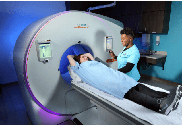
The integration of Positron Emission Tomography (PET) and Computed Tomography (CT) into veterinary research represents a significant advancement in the field. Hybrid PET/CT imaging allows for the simultaneous acquisition of both functional and anatomical data, providing detailed insights into physiological processes within animals. This technology is particularly valuable in understanding complex diseases such as cancer, neurodegenerative disorders, and cardiovascular conditions.
The PET component provides metabolic information, while the CT part offers high-resolution anatomic detail. The combination allows researchers to correlate functional changes with structural alterations, leading to more precise diagnoses and treatment planning. Hybrid PET/CT imaging is not only useful for identifying pathologies but also in monitoring therapeutic efficacy and guiding surgical interventions.
The process typically involves preparing the animal subject according to specific protocols that ensure accurate imaging results. This includes administering radiotracers such as fluorodeoxyglucose (FDG) or other tracers based on the research objectives, which are then imaged using the PET scanner. The CT component provides anatomical context for the functional data acquired by the PET scanner.
The instrumentation used in hybrid PET/CT imaging is highly sophisticated and requires stringent calibration to ensure accuracy and reliability. The equipment must adhere to international standards such as those provided by ASTM, IEC, and ISO to maintain consistency across different facilities. Research institutions often have dedicated laboratories equipped with state-of-the-art scanners that can handle a wide range of animal sizes from small rodents to large dogs.
Reporting the results involves detailed analysis of the images obtained, which is typically performed by experienced radiologists or nuclear medicine specialists who are well-versed in interpreting PET/CT data. This process includes quantifying metabolic activity, identifying areas of abnormal uptake or suppression, and correlating these findings with clinical observations.
| Parameter | Description | Significance |
|---|---|---|
| PET Scan Time | Absorption time of radiotracer in tissues | Directly affects the accuracy of metabolic measurements |
| Anatomic Detail | Resolution provided by CT component | Critical for precise localization and correlation with functional data |
| Radiotracer Uptake | Rate at which radiotracer is absorbed by tissues | Indicates metabolic activity and can be used in diagnostic imaging |
| Image Correlation | Matching of PET and CT images | Enhances the interpretability of functional data within anatomical context |
The application of hybrid PET/CT imaging in veterinary research is broad, ranging from oncology to cardiology. Its ability to provide detailed insights into metabolic processes makes it an indispensable tool for advancing our understanding of diseases and improving treatment strategies.
Industry Applications
- Cancer Research: Identifying tumor characteristics and monitoring response to therapy.
- Disease Modeling: Studying the progression of diseases like Alzheimer’s and Parkinson’s in animal models.
- Veterinary Medicine: Diagnosing and treating complex conditions in pets and livestock.
- Pharmacology: Evaluating drug efficacy and toxicity in preclinical trials.
The versatility of hybrid PET/CT imaging allows it to be applied across various sectors within the healthcare industry, providing valuable data for both basic research and clinical applications.
International Acceptance and Recognition
Hybrid PET/CT imaging has gained widespread acceptance in veterinary research due to its ability to provide comprehensive functional and anatomical insights. The technology is recognized by leading international bodies such as the International Atomic Energy Agency (IAEA), the World Health Organization (WHO), and various national regulatory authorities.
Standards like ISO 5725 for precision and bias of measurement methods, ASTM E1169 for performance specifications of positron emission tomographs, and EN 809 for quality assurance in diagnostic imaging are crucial in ensuring the reliability and accuracy of hybrid PET/CT imaging. Compliance with these standards is essential to maintain high-quality research and diagnostic outcomes.
The use of hybrid PET/CT imaging in veterinary research is supported by numerous scientific publications and clinical guidelines that emphasize its role in advancing knowledge and improving patient care. The technology continues to evolve, driven by ongoing advancements in radiotracer development and scanner design.
Use Cases and Application Examples
Hybrid PET/CT imaging is used in a variety of applications within veterinary research, including:
| Application | Description |
|---|---|
| Cancer Diagnosis | Detecting and characterizing tumors in animals. |
| Tumor Response Monitoring | Evaluating the effectiveness of cancer treatments over time. |
| Neurodegenerative Disease Modeling | Studying the progression of diseases like Alzheimer’s and Parkinson’s in animal models. |
| Veterinary Cardiology | Assessing cardiovascular health and diagnosing conditions such as heart failure. |
| Pharmacokinetics | Evaluating the absorption, distribution, metabolism, and excretion of drugs in animals. |
An example use case involves a study aimed at understanding the progression of Alzheimer’s disease in dogs. Researchers administered radiotracers to dogs exhibiting symptoms of cognitive decline and used hybrid PET/CT imaging to track changes in brain metabolism over time. This provided valuable insights into the disease process and potential therapeutic targets.


