Thoracic Radiography Testing in Veterinary Clinics
The practice of thoracic radiography testing in veterinary clinics is an essential diagnostic tool that provides detailed images of a pet's chest, including the heart, lungs, and other internal structures. This service allows for the early detection and diagnosis of various respiratory conditions, cardiac issues, and other abnormalities within pets. The primary objective is to ensure accurate imaging that can inform necessary medical decisions.
Thoracic radiography in veterinary clinics involves a series of steps aimed at producing high-quality images that are clear enough for detailed analysis by veterinarians and radiologists. Preparation typically includes ensuring the pet remains still during the procedure, which often requires sedation or immobilization for larger animals. The imaging process itself is non-invasive and painless; however, it can be stressful for pets, so proper handling and support are crucial.
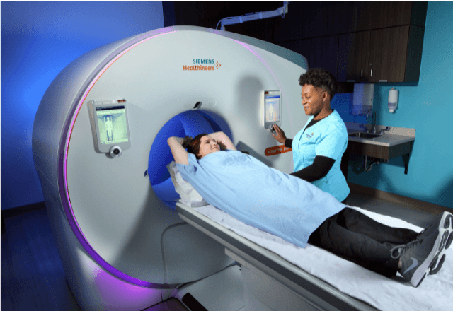
The equipment used in this service consists of a radiographic machine capable of producing low-dose radiation to minimize exposure risks. The images produced are analyzed by trained technicians who ensure the clarity and accuracy of the results. This analysis is then reviewed by veterinarians who use it to develop an appropriate treatment plan or further diagnostic steps.
For accurate thoracic radiography, proper specimen preparation is vital. Pets must be positioned correctly on a radiographic table, often with the help of specialized equipment like immobilization boards for larger animals. The imaging process itself requires minimal movement; even slight shifts can distort the image quality. Once the images are captured, they undergo detailed review and analysis to ensure diagnostic clarity.
Thoracic radiography plays a crucial role in diagnosing respiratory conditions such as pneumonia, bronchitis, and asthma, among others. It is also used for evaluating heart health, detecting tumors or masses within the chest cavity, and identifying foreign bodies or fluid accumulations. The service provides valuable insights into the pet's overall well-being, supporting informed healthcare decisions.
The equipment used in this process includes a radiographic machine capable of producing high-resolution images. This ensures that even subtle changes can be detected early on. Proper calibration and maintenance are critical to maintaining image quality and ensuring consistent results over time.
| Procedure | Details |
|---|---|
| Patient Positioning | The pet is carefully positioned to ensure that the chest area is accessible for clear imaging. This may involve using immobilization boards for larger animals. |
| Radiographic Machine | High-quality, low-dose radiographic machines are used to minimize exposure risks while producing clear images. |
| Image Analysis | Trained technicians analyze the images in detail before they are reviewed by veterinarians for further interpretation and diagnosis. |
The accuracy of thoracic radiography testing is paramount, especially when it comes to identifying issues that require immediate attention. The service ensures that each image captures the necessary details for precise analysis, supporting early intervention in pet health care.
Industry Applications
Thoracic radiography testing has numerous applications across veterinary clinics, from routine check-ups to specialized diagnostic procedures. This section highlights some of the key uses and benefits associated with this service.
| Application | Description |
|---|---|
| Routine Check-Ups | Detecting early signs of respiratory or cardiac issues in pets before they become severe. |
| Respiratory Conditions Diagnosis | Identifying pneumonia, bronchitis, and asthma to ensure timely treatment. |
| Cancer Detection | Detecting tumors or masses within the chest cavity for prompt intervention. |
| Foreign Body Identification | Locating and assessing foreign bodies that may have been ingested by pets. |
| Fluid Accumulation Monitoring | Evaluating fluid build-up in the pleural space or pericardium for appropriate management. |
The versatility of thoracic radiography testing makes it a valuable tool in various diagnostic scenarios, ensuring comprehensive care for pets.
International Acceptance and Recognition
- ISO 15408: This international standard ensures the quality and reliability of radiographic imaging systems used in veterinary clinics.
- ASTM E3697: Provides guidelines for the performance evaluation of radiographic imaging systems, ensuring consistent image quality.
- EN ISO 25587: Sets out specifications for the installation and use of radiographic equipment to ensure safety and effectiveness.
The acceptance of thoracic radiography testing in veterinary clinics is widespread, with these standards being crucial for maintaining the integrity and reliability of diagnostic imaging. Compliance with these guidelines ensures that veterinarians can rely on accurate and consistent results when diagnosing pet health issues.
Environmental and Sustainability Contributions
The practice of thoracic radiography testing in veterinary clinics contributes positively to environmental sustainability by promoting early detection and intervention, which can reduce the need for more invasive diagnostic procedures. By using low-dose radiation techniques and advanced imaging systems, the service minimizes exposure risks while ensuring clear images.
Additionally, proper specimen preparation and equipment calibration contribute to reducing waste and optimizing resource use. The service also supports sustainable healthcare practices by enabling timely treatment plans that can prevent complications and extend pet lifespans.


