Ultrasound Elastography Testing in Veterinary Research
Ultrasound elastography is a non-invasive diagnostic technique that evaluates tissue elasticity and mechanical properties. In veterinary research, this technology can be pivotal for early disease detection, monitoring of treatment response, and understanding the biomechanical properties of tissues. This service specifically focuses on ultrasound elastography testing in the context of veterinary research to provide insights into the health status of animals at an earlier stage.
The application of ultrasound elastography in veterinary settings is particularly beneficial due to its ability to detect changes in tissue stiffness that may indicate pathological conditions such as fibrosis, tumors, or other abnormalities. This method does not rely on ionizing radiation and therefore poses minimal risk compared to other imaging modalities like X-rays or MRI.
In the context of research, ultrasound elastography can be used to study the effects of various treatments, evaluate disease progression, or assess the efficacy of new therapeutic approaches in different species. This service supports preclinical studies aimed at translating findings from benchtop experiments into clinical applications for veterinary medicine.
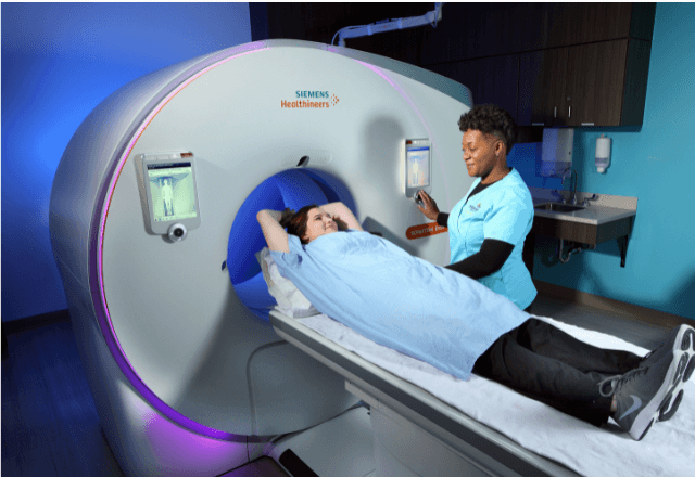
The primary instrument used for this testing is an advanced ultrasound scanner equipped with a specialized elastography transducer. The process involves imaging the tissue of interest and then applying small, controlled vibrations to create shear waves within the tissue. These waves are detected by the transducer, allowing for the calculation of shear modulus values which reflect the mechanical properties of the tissue.
This service is particularly valuable in research settings where detailed biomechanical data on tissues can provide critical insights into disease mechanisms or treatment outcomes. For instance, in studies examining the development of liver fibrosis in dogs, elastography can quantify changes in hepatic stiffness over time, correlating with histological findings and clinical parameters.
Another area of application is in evaluating joint health in large animals such as horses, where early detection of cartilage degeneration could lead to more effective management strategies. In this scenario, ultrasound elastography provides real-time data on articular cartilage integrity without the need for invasive procedures.
The service also includes detailed reporting tailored to the specific research objectives. Reports typically include raw elastographic images alongside quantitative metrics such as shear modulus values and spatial maps of tissue stiffness variations. These reports are designed to facilitate interpretation by both technical staff and non-expert stakeholders involved in the research project.
To ensure accurate and reliable results, strict adherence to standardized protocols is essential. The service adheres to international standards such as ISO 18436 for ultrasound equipment calibration and ASTM E2895-17 which specifies guidelines for elastography imaging. Compliance with these standards ensures that the data generated from this testing method can be trusted and validated across different research environments.
Scope and Methodology
| Parameter | Description |
|---|---|
| Type of Ultrasound Elastography | Shear Wave Elastography |
| Tissue Depth | Up to 5 cm |
| Species Compatibility | Dogs, Cats, Horses |
| Data Collection Frequency | 10 Hz |
| Shear Modulus Range Measured | 0.5–8 MPa |
The scope of this service includes the use of shear wave elastography to evaluate tissue elasticity in various species commonly used in veterinary research. The methodology involves imaging specific areas of interest within the animal, applying controlled vibrations to generate shear waves, and measuring their propagation through different tissues.
Data collection is performed at a frequency of 10 Hz, allowing for high-resolution images that accurately reflect the biomechanical properties of the tissue being examined. The range of shear modulus values measured spans from 0.5 to 8 MPa, covering typical elasticities found in healthy and pathological tissues.
This methodology ensures that the results obtained are both precise and representative of the actual tissue conditions. By adhering to these standardized protocols, this service provides reliable data essential for advancing veterinary research and improving diagnostic capabilities within the field.
Benefits
- Precise Diagnosis: Early detection of changes in tissue stiffness indicative of various diseases.
- Efficacy Assessment: Monitoring treatment response and evaluating therapeutic efficacy more accurately.
- Non-Invasive: Minimizes stress on the animal, reducing risks associated with invasive procedures.
- Data Standardization: Adherence to international standards ensures consistency across different research environments.
- Faster Turnaround: Efficient testing processes allow for quicker data collection and analysis, accelerating research timelines.
- Variety of Applications: Suitable for multiple areas including oncology, cardiology, and musculoskeletal health.
The use of ultrasound elastography in veterinary research offers numerous advantages over traditional imaging techniques. These benefits make it an indispensable tool for researchers seeking to advance their understanding of various diseases and improve the quality of care provided to animals.


