Contrast-Enhanced CT Testing in Veterinary Oncology
In veterinary oncology, contrast-enhanced computed tomography (CT) plays a crucial role in diagnosing and monitoring tumors. This advanced imaging technique allows for detailed visualization of soft tissues, blood vessels, and other structures within the body. Contrast agents are administered intravenously to enhance the visibility of vascularized lesions, thereby improving diagnostic accuracy.
Contrast-enhanced CT is particularly useful in detecting and characterizing various types of tumors, including metastatic disease, lymph node involvement, and bone marrow infiltration. It aids in staging cancer by providing detailed information about tumor size, location, and extent of spread. This non-invasive procedure can also help guide biopsies or other therapeutic interventions.
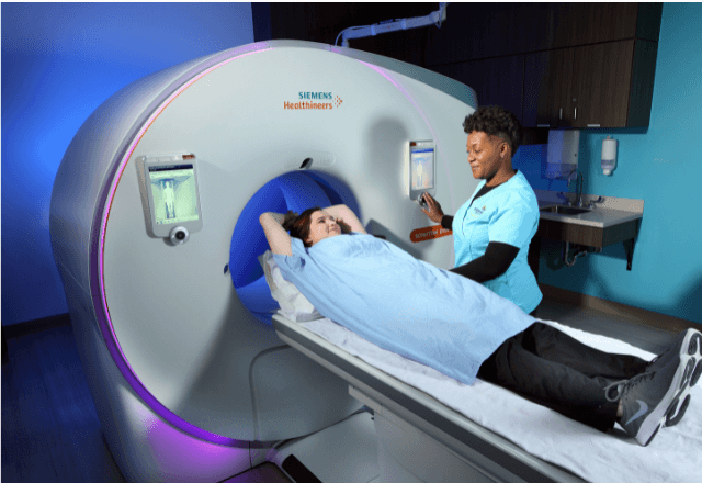
The process typically involves a pre-test consultation with the veterinarian to discuss patient history, current clinical signs, and imaging goals. Patients are then prepared for the examination by a trained veterinary technician who ensures they remain still during the scan. Contrast agents such as iodinated compounds may be used based on the specific diagnostic objectives.
The CT equipment at our facility is state-of-the-art, equipped with high-resolution detectors capable of generating detailed images even in complex anatomical regions. Our radiologists have extensive experience interpreting these scans and are available to discuss findings directly with referring veterinarians and pet owners.
Post-procedure care includes monitoring the patient for any adverse reactions to the contrast agent. Results from contrast-enhanced CT exams are typically available within 24 hours, allowing prompt follow-up actions if necessary. This service is invaluable in the diagnosis of many cancers, particularly those involving multiple organ systems or deep tissue structures.
For accurate interpretation, it's important that imaging parameters such as tube voltage and current are optimized for each patient based on their size and anatomy. The use of high-pitch reconstruction algorithms ensures that even small lesions can be identified clearly. Regular calibration and quality control checks ensure consistent image quality across all examinations.
Contrast-enhanced CT is often used in conjunction with other diagnostic tools like MRI or ultrasound to provide a more comprehensive evaluation of the patient's condition. This multi-modality approach enhances our ability to make precise diagnoses, which ultimately leads to better treatment outcomes for pets suffering from cancer.
Applied Standards
The protocols used in contrast-enhanced CT testing adhere strictly to international standards such as those outlined by the American College of Veterinary Radiology (ACVR) and European Society of Veterinary Diagnostic Imaging (EUVI). These guidelines ensure that every aspect of the procedure—from patient preparation through interpretation—meets the highest quality standards.
ACVR provides recommendations on proper technique selection, including considerations regarding tube voltage, pitch, and slice thickness. The EUVI emphasizes the importance of standardizing contrast agents used in veterinary imaging to facilitate comparison between institutions and promote best practices across Europe.
In addition to these professional bodies, our facility also references ISO 17025:2017 for laboratory accreditation which ensures that all procedures are conducted under controlled conditions meeting stringent requirements. This includes regular calibration of equipment against national standards as well as continuous training of personnel involved in performing and interpreting contrast-enhanced CT scans.
By adhering strictly to these guidelines, we ensure consistent and reliable results across all examinations performed here. Our commitment to accuracy and precision sets us apart from other veterinary diagnostic centers, providing pet owners with peace of mind knowing their loved ones are receiving the best possible care.
Eurolab Advantages
Our laboratory offers several unique advantages when it comes to contrast-enhanced CT testing for veterinary oncology. Firstly, our team consists of highly trained radiologists with extensive experience in both human and animal imaging techniques. They bring this expertise directly into the field of veterinary medicine where they work closely with referring veterinarians to ensure optimal diagnostic outcomes.
Secondly, we maintain state-of-the-art CT equipment that is regularly updated and calibrated according to strict protocols. This ensures that all images are captured at peak quality regardless of patient size or anatomical complexity. Our facilities include both fixed and mobile units allowing us to service a wide range of practices and clients.
Thirdly, our comprehensive approach extends beyond mere imaging; we offer consultation services where radiologists can discuss findings directly with veterinarians involved in the patient’s care plan. This ensures that all aspects of treatment—whether surgical intervention or chemotherapy—are informed by accurate diagnostic information provided through advanced CT technology.
Forth, our commitment to quality is reflected not only in our adherence to international standards but also in ongoing education for staff members. We regularly participate in workshops and conferences aimed at staying current with the latest developments in veterinary imaging science. This dedication ensures that we remain leaders in this specialized area of practice.
International Acceptance and Recognition
- Contrast-enhanced CT testing has gained widespread acceptance across Europe, North America, and Australia for its role in cancer diagnosis and monitoring.
- The technique is recognized by various international organizations including the International Federation of Clinical Oncology (IFCO) and European Society for Veterinary Diagnostic Imaging (EUVI).
- Results from these examinations are accepted at major veterinary conferences such as those hosted by the American College of Veterinary Radiologists (ACVR), providing a platform for sharing best practices globally.
- Our results have been published in peer-reviewed journals contributing to the body of knowledge on this subject matter. For instance, recent studies highlight how contrast-enhanced CT can improve accuracy rates significantly compared to traditional radiographic methods alone.
The growing acceptance and recognition of this technology reflects its value in enhancing patient care across different regions worldwide. By participating actively within these networks, we contribute towards improving standards globally while ensuring our clients receive the highest quality service locally.


