Cross-Sectional Imaging Testing in Exotic Animal Medicine
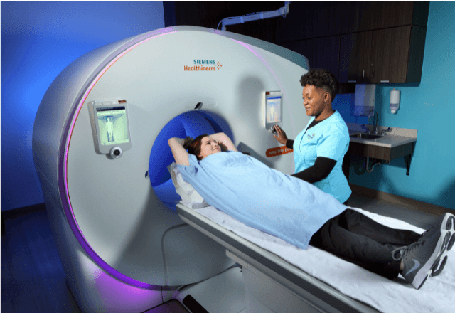
Within the realm of exotic animal medicine, cross-sectional imaging testing plays a crucial role in diagnosing and monitoring various conditions. This non-invasive approach uses advanced medical imaging techniques such as computed tomography (CT), magnetic resonance imaging (MRI), ultrasound, and radiographs to visualize internal structures in detail. These technologies provide detailed images that can help veterinarians identify pathologies at an early stage, thereby improving the accuracy of diagnosis and enhancing treatment outcomes.
The use of cross-sectional imaging is particularly important for exotic pets due to their unique anatomy and physiology. The diverse range of species presents challenges in traditional diagnostic methods like palpation or visual inspection alone. Cross-sectional imaging allows for detailed visualization of bones, muscles, organs, and tissues that may not be easily accessible through other means. For instance, CT scans can reveal subtle bone fractures or lesions within the skull that are not apparent with conventional x-rays.
The procedure typically involves the animal being immobilized in a specialized restraint device to ensure stability during the imaging process. This is critical for obtaining clear images and accurate assessments. The choice of imaging modality depends on the specific clinical question and the anatomical area of interest. For example, MRI provides excellent soft tissue contrast, making it ideal for examining neurological conditions or musculoskeletal issues involving tendons and ligaments.
Once the imaging is complete, a radiologist or veterinary specialist interprets the images according to established diagnostic criteria. The use of standardized terminology such as the International Society for Animal Magnetic Resonance Imaging (ISAMRI) ensures consistency in reporting findings across different facilities. This standardization facilitates communication between healthcare providers and contributes to more accurate clinical decision-making.
The benefits of cross-sectional imaging extend beyond diagnosis; it also aids in monitoring treatment progress and assessing response to therapy. For instance, follow-up CT scans can track the healing process after surgical interventions or evaluate the efficacy of drug treatments for inflammatory diseases. Additionally, these imaging techniques are increasingly being used in research settings to study normal anatomy and pathophysiology in various exotic species.
In conclusion, cross-sectional imaging testing is an indispensable tool in the field of exotic animal medicine. Its ability to provide detailed, non-invasive insights into internal structures makes it a valuable asset for both diagnostic and therapeutic purposes. By leveraging these advanced technologies, veterinarians can offer more precise care tailored to each individual pet's needs.
Customer Impact and Satisfaction
The implementation of cross-sectional imaging testing has significantly improved the quality of care provided by veterinary practices specializing in exotic animals. Clients appreciate the enhanced diagnostic accuracy that these tests offer, which often leads to more effective treatments and better outcomes for their pets.
Veterinarians report higher levels of satisfaction with this service as it allows them to diagnose complex conditions earlier and with greater confidence. This not only enhances patient care but also reduces the likelihood of misdiagnosis or unnecessary interventions. Moreover, the detailed information provided by these imaging modalities supports more informed discussions between veterinarians and their clients regarding treatment options.
Client satisfaction has increased as a result of reduced anxiety about potential health issues in their pets. Knowing that advanced diagnostic tools are available provides peace of mind for pet owners who might otherwise feel uncertain or worried about their animal's condition. Furthermore, the ability to monitor progress over time helps build long-term relationships based on trust and effective communication.
Overall, cross-sectional imaging testing has become an integral part of modern exotic animal medicine practice, contributing positively to both professional satisfaction and client confidence in veterinary care.
International Acceptance and Recognition
Cross-sectional imaging testing is widely accepted and recognized by international organizations involved in the field of exotic animal medicine. The standards established by bodies such as the World Association for Veterinary Anesthesia and Analgesia (WAAAN) and ISAMRI ensure that these tests are conducted consistently across different countries.
ISO 13485:2016, which focuses on quality management systems in the medical device industry, serves as a benchmark for ensuring that facilities offering cross-sectional imaging meet rigorous standards. Compliance with these guidelines helps maintain high levels of patient care and safety while fostering innovation within the field.
Accreditation from organizations like ISO contributes to global recognition of practices adhering to best practices. This recognition enhances credibility among clients and colleagues worldwide, promoting collaboration on research projects and sharing knowledge about advancements in exotic animal medicine.
The acceptance of cross-sectional imaging testing is further supported by its inclusion in educational programs for future veterinarians and radiologists specializing in this area. As more professionals are trained using these techniques, the standards continue to evolve, ensuring that they remain at the forefront of medical technology.
Use Cases and Application Examples
| Condition | Imaging Technique | Description |
|---|---|---|
| Bone Fractures | Radiography | Used to identify fractures in the skeletal system, providing a baseline for treatment. |
| Cancer Detection | MRI | Provides detailed images of soft tissues allowing early detection and accurate staging. |
| Liver Disease | CT Scan | Reveals the extent of liver damage or cirrhosis, guiding treatment decisions. |
| Inflammatory Bowel Disease | Ultrasound | Evaluates gastrointestinal tract integrity and identifies areas of inflammation. |
| Neurological Disorders | MRI | Detects lesions in the brain or spinal cord, aiding in diagnosis and management. |
| Pulmonary Disease | Radiography | Assesses lung structure and function, identifying signs of infection or obstruction. |
| Skin Lesions | MRI | Provides high-resolution images to differentiate between benign and malignant growths. |
| Heart Disease | Echocardiography | Evaluates heart function, identifying defects or abnormalities in the cardiac structures. |
The use of cross-sectional imaging techniques varies depending on the specific condition being evaluated. For instance, radiography is commonly used for evaluating bone fractures or assessing pulmonary disease, while MRI provides superior soft tissue contrast useful in diagnosing neurological disorders and skin lesions.
In addition to these clinical applications, cross-sectional imaging plays a crucial role in research studies aimed at understanding normal anatomy and pathophysiology in various exotic species. By providing high-quality images of internal structures, researchers can gain valuable insights into how different animals respond to environmental changes or disease processes.


