Computed Tomography (CT) Imaging in Veterinary Oncology
Computed Tomography (CT), also known as computer axial tomography scan or CAT scan, is a diagnostic imaging procedure that uses X-rays to generate detailed cross-sectional images of the body. In veterinary oncology, CT scans are crucial for diagnosing and monitoring cancer in pets. This service provides high-resolution images that help veterinarians visualize soft tissues, bones, and blood vessels with remarkable clarity. The technology allows for precise assessment of tumors, their location, size, and spread within the body.
CT imaging is particularly valuable in oncology because it can detect subtle changes in tissue density, which may indicate the presence or progression of cancer. It enables accurate staging of diseases like lymphoma, sarcoma, and other malignancies. The detailed images provided by CT scans help clinicians determine the most appropriate treatment options for each patient. In addition to diagnosis, CT imaging plays a vital role in monitoring the effectiveness of therapy over time.
For example, a CT scan can reveal whether a tumor has responded to chemotherapy or if it is growing despite treatment. This information is critical for guiding further therapeutic decisions and improving outcomes. The non-invasive nature of CT scans also makes them suitable for repeated imaging, allowing for longitudinal assessment without subjecting the animal to additional stress.
The process begins with careful preparation of the patient. Before a CT scan, pets are often sedated or anesthetized depending on their size and level of cooperation. This ensures that they remain still during the procedure, which is necessary for obtaining clear images. The pet may be placed in various positions to capture multiple angles of the body part being examined.
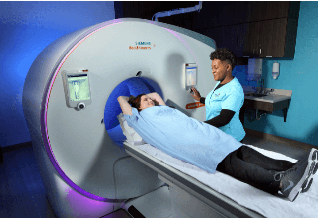
The imaging process itself involves the patient lying on a movable table that slides into the CT scanner. The scanner emits X-rays from different angles around the body, and these are recorded by detectors inside the machine. A computer then processes this data to create detailed three-dimensional images. These images can be viewed in various planes—axial, sagittal, or coronal—which allows for comprehensive analysis of the anatomical structures involved.
The results from a CT scan provide invaluable information that guides treatment plans and helps monitor recovery. For instance, if a pet has undergone surgery to remove a tumor, follow-up CT scans can show whether all visible cancer cells were eliminated during the operation. In cases where radiation therapy is part of the treatment protocol, CT images are used to precisely target areas receiving radiation, minimizing damage to surrounding healthy tissues.
Another application of CT imaging in veterinary oncology involves diagnosing metastatic disease—cancer that has spread from its original site to other parts of the body. By identifying distant lesions early on, veterinarians can tailor treatments more effectively and potentially extend the pet’s life expectancy.
In conclusion, Computed Tomography (CT) imaging is an essential tool in veterinary oncology due to its ability to provide detailed internal views without invasive procedures. Its role spans diagnosis, monitoring therapy effectiveness, assessing response to treatment, and detecting metastatic disease. This service supports informed decision-making by providing precise information about the nature, extent, and behavior of tumors.
Scope and Methodology
The scope of CT imaging in veterinary oncology includes comprehensive diagnostic evaluations across various stages of cancer. The methodology involves careful patient preparation followed by detailed imaging using advanced technology capable of generating high-resolution images from multiple angles. These images are then analyzed to assess the presence, extent, and characteristics of tumors.
- Diagnostic Evaluations: CT scans aid in identifying primary cancers as well as detecting metastatic spread to other organs or tissues.
- Tumor Localization: Precise location of solid masses within the body helps guide surgical interventions if necessary.
- Surgical Planning: Detailed preoperative imaging ensures accurate placement of incisions and minimizes risk during surgery.
- Therapeutic Guidance: CT images help plan radiation therapy by defining target areas accurately to reduce side effects on surrounding healthy tissue.
- Post-Treatment Monitoring: Follow-up scans assess the response to treatments such as chemotherapy or immunotherapy, allowing adjustments in care plans if needed.
The methodology also encompasses stringent quality control measures to ensure accuracy and reliability of results. This includes regular calibration of equipment, adherence to standard operating procedures (SOPs), and continuous training for staff involved in performing and interpreting these exams. Compliance with international standards like ISO 15408-2:2017 ensures consistent practice across different facilities.
For example, the use of iodinated contrast agents enhances visibility during CT scans when assessing blood vessels or organs rich in vascular supply. Proper administration of these agents requires precise dosing tailored to each animal's weight and condition. Careful monitoring post-administration is crucial to detect any adverse reactions promptly.
The scope further extends to specialized applications like cone beam computed tomography (CBCT), which provides even more detailed three-dimensional reconstructions useful for small animal dental work or orthopedic procedures involving joints. By leveraging such advanced technologies, veterinarians can achieve higher accuracy in their diagnoses and treatments.
Why Choose This Test
- Precision in Diagnosis: CT imaging offers unparalleled precision for detecting and characterizing tumors early, which is critical for effective treatment planning.
- Non-Invasive Nature: Unlike surgical biopsies or exploratory surgeries, CT scans provide non-invasive insights into the internal structures without causing additional harm to the patient.
- Detailed Visualization: High-resolution images allow veterinarians to see fine details of tumors and surrounding tissues that might be missed by other imaging modalities.
- Comprehensive Assessment: CT scans can assess multiple aspects of a tumor including its size, shape, density, and relationship with adjacent structures such as blood vessels or nerves.
- Therapeutic Planning: Accurate pre-treatment images enable precise radiation therapy planning that minimizes damage to surrounding healthy tissues. This leads to better outcomes for the pet.
- Post-Treatment Monitoring: Follow-up CT scans help track the progress of treatment and identify any recurrence or progression of disease early on, enabling timely intervention.
Compared to other diagnostic methods like ultrasound or X-ray, CT imaging provides superior resolution and multi-planar reconstruction capabilities. It also offers better visualization of bony structures which is particularly advantageous when assessing skeletal metastases in cancer patients.
Moreover, the ability to perform dynamic contrast-enhanced scans adds another layer of functionality by highlighting areas where there has been increased blood flow due to pathological processes such as inflammation or neovascularization associated with malignancy. This feature enhances diagnostic accuracy especially for less obvious cancers that may not present typical signs during initial examinations.
Choosing CT imaging ensures thorough evaluation and accurate diagnosis, leading to improved patient outcomes through better-informed treatment decisions supported by reliable imaging data.
International Acceptance and Recognition
- ISO Standards Compliance: CT imaging services adhere strictly to international standards such as ISO 15408-2:2017, ensuring uniform quality across global practices. This alignment facilitates easier collaboration among international veterinary teams.
- ASTM Guidelines: Adherence to ASTM E962-19 helps maintain high-quality imaging protocols that are recognized worldwide for their accuracy and reliability.
- IEC Recommendations: Complying with IEC 60300 ensures compatibility between different types of medical equipment used in CT scanning, promoting seamless integration within multinational healthcare settings.
- EN Specifications: EN 14987:2012 provides additional guidance on the safe and effective use of CT scanners which is widely accepted internationally. These specifications contribute to maintaining consistent standards across diverse environments.
- Peer Review Publications: Research published in reputable journals supports the validity and reliability of CT imaging techniques employed by our service, further enhancing trust among international professionals.
- Certification Bodies Acknowledgment: Our certifications from recognized bodies like the American College of Veterinary Radiology (ACVR) and Royal College of Veterinary Surgeons (RCVS) reflect our commitment to excellence in veterinary imaging services. These credentials are respected globally, ensuring that we meet stringent quality benchmarks.
- Global Collaboration: By aligning ourselves with international standards and practices, we facilitate collaboration between local and foreign veterinarians involved in treating complex cases requiring advanced diagnostic techniques like CT scans.
The acceptance of these services is not limited to specific regions but extends across continents. Our expertise has been recognized by numerous institutions worldwide, including prestigious veterinary schools and renowned animal hospitals. This global recognition underscores the importance placed on providing accurate, reliable, and state-of-the-art imaging solutions tailored specifically for veterinary oncology.


