Abdominal Radiography Testing in Livestock
In the clinical and healthcare sector, particularly within radiology testing, abdominal radiography plays a crucial role in diagnosing various internal conditions that affect livestock. This non-invasive diagnostic technique is essential for identifying pathologies such as gastrointestinal obstructions, urolithiasis, pneumonia, and other internal injuries or infections. The primary goal of this service is to ensure the health and welfare of animals by providing accurate diagnostic information without causing additional stress.
Abdominal radiography involves taking X-ray images of an animal's abdominal cavity using specialized equipment designed for veterinary use. This method allows for a detailed examination of the organs, tissues, and structures within the abdomen. The service is particularly valuable in livestock management as it can help detect early signs of disease or injury that might otherwise go unnoticed.
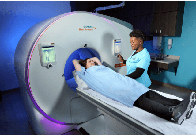
The process begins with thorough preparation to ensure clear images. Animals are carefully positioned on an imaging table, often under light sedation if necessary, to maintain stillness during exposure. The radiographer then selects the appropriate settings for the size and type of animal being examined. The use of high-quality film or digital detectors ensures that even subtle changes in density can be detected.
Once the images are captured, they are analyzed by a qualified veterinarian who interprets the findings according to international standards such as ISO 5210-1:2019 and ASTM E687-14. The radiologist then provides a detailed report outlining any abnormalities observed along with recommendations for further action or treatment.
The importance of this service cannot be overstated, especially in large-scale farming operations where the health of thousands of animals can impact overall productivity and profitability. Early detection through abdominal radiography helps prevent the spread of disease within herds and reduces the need for more invasive diagnostic procedures later on. Moreover, it supports sustainable agricultural practices by minimizing unnecessary interventions and promoting animal welfare.
For quality managers responsible for ensuring operational standards across farms, incorporating regular abdominal imaging into routine health checks can significantly improve herd health metrics. Compliance officers will appreciate how this service aligns with regulatory requirements aimed at maintaining high ethical standards in livestock care. R&D engineers may find value in understanding the technical aspects of radiography equipment and its applications beyond standard use cases.
When selecting a laboratory for abdominal radiography testing, look for facilities that adhere strictly to best practices and employ experienced personnel trained specifically in veterinary diagnostic imaging techniques.
Why It Matters
Regular abdominal radiography is vital for maintaining the health of livestock herds. By identifying issues early on, farmers can take proactive steps to address potential problems before they escalate into larger concerns that could affect entire populations. This preventive measure helps reduce mortality rates among animals and ensures a more productive workforce capable of performing optimally.
From an economic perspective, preventing costly losses due to untreated health conditions translates directly into increased profitability for farms. Additionally, improved animal welfare contributes positively towards public perception regarding the ethical treatment of animals in agriculture. It also enhances biosecurity measures by reducing the risk of disease outbreaks within and between herds.
- Reduces mortality rates among livestock
- Improves overall herd health metrics
- Promotes sustainable agricultural practices
- Enhances biosecurity through early detection
- Maintains high ethical standards in animal care
The ability to diagnose internal conditions accurately and promptly is crucial for effective management of livestock. Farmers who invest time and resources into this service are likely to see significant returns not only financially but also environmentally by minimizing waste and promoting healthier ecosystems.
Scope and Methodology
| Step | Description |
|---|---|
| Preparation | The animal is gently positioned on the imaging table. If necessary, light sedation may be administered to keep the subject still during exposure. |
| Selecting Settings | The radiographer adjusts the settings based on the size and type of the animal being examined to ensure optimal image quality. |
| Exposure | X-ray images are captured using specialized veterinary equipment designed for this purpose. High-quality film or digital detectors are used depending upon preference. |
| Analysis | A qualified veterinarian analyzes the images and provides a detailed report on any abnormalities observed along with appropriate recommendations. |
The methodology employed ensures that each step is performed meticulously to achieve reliable diagnostic results. Compliance with international standards like ISO 5210-1:2019 guarantees consistency in the quality of service provided across different locations.
Environmental and Sustainability Contributions
- Reduces the need for invasive diagnostic procedures through early detection
- Promotes sustainable farming practices by minimizing unnecessary interventions
- Supports animal welfare initiatives aimed at enhancing overall health
- Aids in maintaining biosecurity measures within herds and between them
- Contributes to better public perception regarding ethical treatment of animals in agriculture
The environmental impact of this service is minimal compared to the benefits it brings. By catching problems early, less medicine needs to be used, reducing antibiotic resistance concerns. Moreover, fewer sick animals mean reduced methane emissions from stress-induced behaviors.


