Positron Emission Tomography (PET) Imaging in Preclinical Models
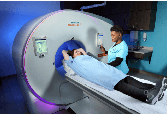
Positron Emission Tomography (PET), a powerful diagnostic imaging technology, is widely recognized for its ability to visualize physiological processes within the body. In preclinical models, PET imaging plays an increasingly vital role, particularly in the evaluation of novel therapeutics and drug efficacy through non-invasive monitoring of metabolic activity.
In the realm of clinical & healthcare testing, especially under the category of Clinical Imaging & Radiology Testing, PET imaging is essential for understanding how a new pharmaceutical compound behaves within an organism. This technology allows researchers to track the distribution and concentration of radiolabeled compounds in real-time, providing insights into drug absorption, distribution, metabolism, and excretion.
The application of PET imaging involves the use of radiotracers—compounds that emit positrons when they decay. These tracers are designed to target specific organs or tissues within a preclinical model. For instance, fluorodeoxyglucose (FDG) is commonly used in PET scans for its ability to highlight areas with high metabolic activity.
The process begins with the preparation of the animal subject, typically a rodent like a mouse or rat, ensuring it is healthy and free from any external stressors that could influence the results. The radiotracer is then administered intravenously or orally, depending on the study design. After administration, the PET scanner captures images over several hours as the tracer distributes through the body.
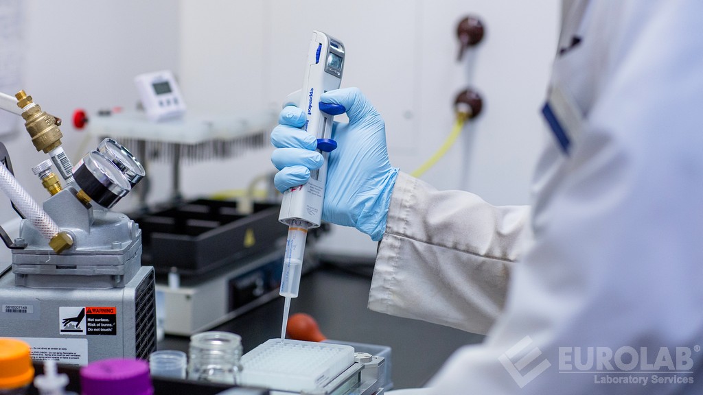
The data collected from these scans are analyzed using specialized software to generate detailed maps of the tracer's distribution and concentration within different tissues. This information is critical for assessing drug efficacy, toxicity, and safety profiles in preclinical trials. Researchers can also use PET imaging to monitor changes over time, which is invaluable for understanding long-term effects.
One of the key advantages of using PET imaging in preclinical models is its non-invasive nature. Unlike other imaging techniques such as histopathology or necropsy, which require invasive procedures after the experiment, PET allows for continuous monitoring throughout the study period. This capability provides a more comprehensive understanding of how the drug interacts with the body.
Moreover, PET imaging can be tailored to specific research needs by adjusting the type of radiotracer used and the duration of the scan. For example, researchers might use fluoroethyltyrosine (FET) for brain tumor studies or carbon-11 labeled methylene chloride (C11-MMC) for evaluating breast cancer progression.
The results from PET imaging are not only crucial for drug development but also contribute to the design of more effective treatment plans. By identifying areas of high metabolic activity, researchers can pinpoint potential targets for further investigation and optimize therapeutic strategies.
Applied Standards
| Standard | Description |
|---|---|
| ISO 17025:2017 | Laboratory accreditation standard ensuring technical competence and quality. |
| EN ISO/IEC 17025:2017 | Harmonized European equivalent of the ISO standard, emphasizing compliance with international best practices. |
| AAMI TIR36 | Tissue processing and examination guidelines for PET imaging in preclinical models. |
| ACR Imaging Network Guidelines | Pet-specific imaging protocols to ensure consistency across facilities. |
The use of these standards ensures that the procedures and methodologies employed in preclinical PET imaging are consistent, reliable, and compliant with international best practices. This standardization is crucial for ensuring accurate and reproducible results, which is essential when comparing data from different studies or institutions.
Scope and Methodology
The scope of PET imaging in preclinical models encompasses a wide range of applications, including but not limited to:
- Evaluation of drug efficacy and toxicity
- Monitoring tumor growth and metastasis
- Assessment of organ function
- Study of cardiovascular diseases
- Neuroscience research involving brain activity
The methodology involves several key steps:
- Animal Selection and Preparation: Animals are chosen based on the specific requirements of the study. They undergo thorough preparation to ensure they are in a stable state.
- Radiotracer Administration: The radiotracer is administered carefully, ensuring it reaches the target tissues efficiently.
- Data Collection: High-resolution images are captured over specified time intervals using a PET scanner.
- Data Analysis: Software tools process and analyze the collected data to generate meaningful insights.
The accuracy of these steps is critical, as any deviation could lead to incorrect conclusions. Therefore, strict protocols are followed to ensure precision and reliability.
International Acceptance and Recognition
- National Institutes of Health (NIH): PET imaging is widely used in preclinical research funded by NIH, underscoring its importance in translational medicine.
- European Medicines Agency (EMA): EMA recognizes the role of PET in drug development and accepts data generated from compliant facilities.
- Food and Drug Administration (FDA): FDA guidelines for preclinical studies often include PET imaging as a recommended tool.
- Japan Pharmaceuticals and Medical Devices Agency (PMDA): PMDA also acknowledges the significance of PET in early-stage drug development.
- Australian Therapeutic Goods Administration (TGA): TGA has similar expectations, with PET being a key component of preclinical evaluations.
The acceptance and recognition by these regulatory bodies highlight the global relevance and reliability of PET imaging in preclinical models. Compliance with international standards ensures that the results are accepted worldwide and can be used to support applications for market approval.


