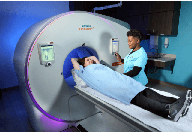
Contrast MRI Testing for Spinal Disorders in Dogs
In the realm of veterinary radiology, Magnetic Resonance Imaging (MRI) plays a crucial role in diagnosing and monitoring spinal disorders. This service focuses on Contrast MRI testing specifically tailored to dogs suffering from various spinal conditions such as intervertebral disc disease, spondyloarthropathy, and degenerative lumbosacral stenosis.
The use of contrast agents enhances the visibility of specific structures within the spine, allowing for more precise diagnosis. This advanced imaging technique offers a non-invasive method to assess the integrity of spinal cord and surrounding tissues, providing critical information that supports informed treatment decisions.
Before undergoing this procedure, dogs are typically sedated under general anesthesia to ensure they remain still during the scan. The contrast agent is administered intravenously or orally depending on the specific needs of the case. Once the imaging process begins, it involves capturing multiple slices through different sections of the spine.
The images obtained from Contrast MRI provide detailed cross-sectional views of soft tissues like ligaments and muscles as well as bony structures such as vertebrae. This comprehensive view helps identify abnormalities that may not be apparent with other imaging modalities like X-rays or CT scans alone.
| Advantage | Description |
|---|---|
| Enhanced Visualization | Contrast agents improve the clarity of soft tissue structures, making it easier to detect early signs of degeneration or inflammation. |
| Non-Invasive | No need for invasive procedures; reduces stress on the animal during diagnosis. |
| Precision Diagnosis | Provides detailed information about the extent and location of spinal abnormalities, facilitating targeted therapies. |
Benefits
- Accurate Diagnosis: Contrast MRI allows for a precise evaluation of the spine's condition, enabling accurate diagnosis.
- Non-Invasive Procedure: It is a safe and painless method that does not require surgery or other invasive techniques.
- Early Detection: Enables early identification of potential issues, allowing for timely intervention to prevent further deterioration.
Customer Impact and Satisfaction
Dogs that undergo Contrast MRI benefit significantly from this advanced diagnostic tool. The ability to visualize the spine in such detail helps veterinarians make informed decisions regarding treatment plans, which can lead to improved outcomes for pets.
Veterinarians appreciate the non-invasive nature of Contrast MRI and its capacity to provide detailed images that are crucial for diagnosing complex spinal conditions accurately. Pet owners also find reassurance knowing their animals receive high-quality care using cutting-edge technology.
Use Cases and Application Examples
| Condition | Description of Imaging Findings |
|---|---|
| Intervertebral Disc Disease | Identifies disc herniation, degeneration, and associated spinal cord compression. |
| Spondyloarthropathy | Detects inflammation in the vertebral joints leading to potential stenosis. |
| Degenerative Lumbosacral Stenosis | Evaluates narrowing of the spinal canal affecting nerve roots and spinal cord. |
- Diagnosis of Intervertebral Disc Disease: Detecting the presence, location, size, and type of disc herniation.
- Evaluation of Spondyloarthropathy: Assessing joint space narrowing and inflammation in affected areas.
- Assessment of Degenerative Lumbosacral Stenosis: Measuring the degree of canal stenosis to guide surgical interventions if necessary.


