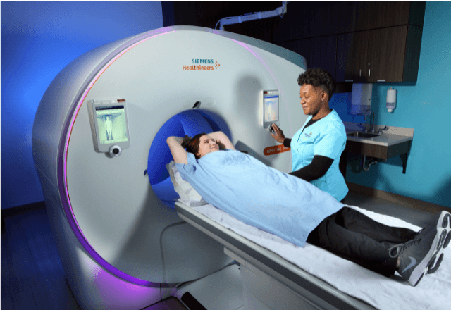
Digital Radiography Testing in Small Animal Clinics
Digital radiography testing has revolutionized diagnostic imaging in small animal clinics. This advanced technology allows veterinarians to capture high-resolution images of a pet's internal anatomy, aiding in the accurate diagnosis and treatment planning for various medical conditions. The process involves exposing an X-ray beam to the subject (typically a small animal) and recording the resulting image on a digital detector rather than photographic film.
The use of digital radiography offers several advantages over traditional film-based methods. Firstly, it provides immediate results, allowing veterinarians to make quick decisions about further treatment or discharge. Secondly, digital images can be easily stored, shared, and manipulated using software tools, enhancing diagnostic capabilities. Thirdly, the technology supports a wide range of imaging protocols tailored to small animal anatomy.
The digital radiography process typically involves placing the small animal on a specialized examination table with its body part aligned correctly for optimal image capture. A technologist or veterinarian then positions and secures the pet before taking X-ray images using a digital detector placed in contact with the skin. After capturing the necessary images, the data is transferred to a computer system where it can be processed and analyzed.
For quality assurance, strict adherence to standard protocols ensures consistent image quality and diagnostic accuracy. This includes calibrating the equipment regularly, maintaining proper exposure settings, and ensuring that personnel are trained in digital radiography techniques. The use of appropriate shielding materials is also crucial for minimizing radiation exposure to both animals and humans.
The primary goal of digital radiography testing in small animal clinics is to provide accurate diagnostic information swiftly. By leveraging this technology, veterinarians can enhance patient care by identifying issues early, which often leads to better outcomes. Additionally, the availability of high-quality images supports more precise surgical planning when necessary.
Standards such as ISO 15343-2:2007 specify requirements for digital radiography systems used in veterinary medicine, ensuring that these devices meet strict quality and performance criteria. Compliance with these standards is essential to guarantee the reliability of diagnostic images produced by the equipment.
Applied Standards
| Standard Code | Description |
|---|---|
| ISO 15343-2:2007 | Specifications for digital radiography systems in veterinary medicine. |
| AAMI PB26:2019 | Performance criteria for mammography imaging systems, which can be relevant to certain types of small animal radiographic examinations. |
Benefits
- Immediate image availability, enabling faster diagnosis and treatment.
- Ease of storage and sharing of digital images across platforms.
- Enhanced diagnostic capabilities through advanced software tools.
- Potential reduction in radiation exposure compared to analog methods when used correctly.
Use Cases and Application Examples
- Chest X-rays for assessing heart size, lung conditions, and presence of foreign bodies.
- Bone fractures and joint injuries evaluation for diagnosis and surgical planning.
- Abdominal imaging to detect organ enlargement, masses, or other abnormalities.


