Orthopedic Radiology Testing in Horses
Orthopedic radiology testing in horses is a critical component of equine healthcare that involves the use of advanced imaging technologies to evaluate and diagnose orthopedic conditions. This service focuses on providing detailed images of the horse's bones, joints, soft tissues, and other anatomical structures using various modalities such as X-rays, computed tomography (CT), magnetic resonance imaging (MRI), and ultrasound.
The primary objective of this testing is to identify, evaluate, and monitor orthopedic issues that can affect a horse's mobility and overall health. Common conditions include fractures, ligament tears, joint disorders, arthritis, and developmental bone problems in growing foals. By identifying these issues early, veterinarians and equine specialists can develop appropriate treatment plans, which may range from conservative management to surgical interventions.
Imaging modalities used in this testing are selected based on the specific anatomical area of interest and the nature of the suspected condition. For instance:
- X-rays: Provide two-dimensional images that can reveal fractures, dislocations, and joint space changes. This is often the first imaging modality used for initial screening.
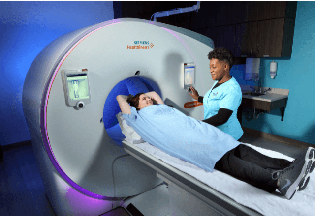
- CT Scans: Offer three-dimensional reconstructions of bones with high resolution. CT scans are particularly useful in evaluating complex fractures or assessing internal structures not easily seen on X-rays.
- MRI Imaging: Capable of providing detailed images of soft tissues such as ligaments, tendons, and cartilage. This is essential for diagnosing conditions like cruciate ligament tears or meniscal injuries in horses.
- Ultrasound: Useful for evaluating superficial structures such as muscles, tendons, and ligaments. It can also be used to assess blood flow and tissue integrity during lameness evaluations.
The process of orthopedic radiology testing begins with a thorough clinical examination conducted by an equine veterinarian or specialist. This examination helps determine the most appropriate imaging modality and aids in interpreting the results once images are obtained. Preparing the horse for the test involves ensuring it is stable on its feet, which may require sedation if necessary to obtain clear images.
Once the horse is positioned appropriately, the imaging equipment is calibrated according to manufacturer specifications and international standards such as ISO 15476-2:2013 (X-ray equipment for radiography of small animals). The operator ensures that all safety measures are in place before starting the procedure. Post-exposure, images are reviewed by a board-certified veterinary radiologist who provides a detailed report outlining findings and recommendations.
The comprehensive nature of orthopedic radiology testing allows for precise diagnosis and tailored treatment plans. This service is not only essential for managing existing conditions but also plays a crucial role in preventive care by identifying potential issues early on. In summary, orthopedic radiology testing in horses represents a vital tool in the equine healthcare toolkit, emphasizing precision, reliability, and non-invasive diagnostic capabilities.
Why It Matters
Orthopedic radiology testing is indispensable for the diagnosis and management of musculoskeletal disorders in horses. These conditions can significantly impact a horse's performance and quality of life, leading to reduced productivity and increased risk of injury if left untreated. Early detection through advanced imaging techniques enables veterinarians to implement timely interventions that improve outcomes.
The importance of this service is underscored by the diverse range of orthopedic issues it addresses:
- Fractures: Accurate identification and staging of fractures are crucial for proper treatment planning. Imaging helps determine the location, type, and severity of the fracture.
- Ligament Injuries: Detailed imaging can differentiate between partial and complete tears, guiding the choice between conservative therapy or surgical repair.
- Joint Disorders: Early recognition of joint degeneration aids in managing osteoarthritis effectively. This includes monitoring progression over time and adjusting treatment strategies accordingly.
- Developmental Bone Problems: Detecting issues during critical growth periods ensures appropriate interventions to prevent long-term disabilities.
In addition to clinical benefits, the use of advanced imaging technologies also enhances patient safety by minimizing the need for invasive procedures. This non-invasive approach is particularly beneficial in equine patients where restraint can be challenging.
The financial implications of untreated orthopedic conditions should not be overlooked. Chronic pain and lameness can lead to prolonged recovery times, increased veterinary costs, and diminished earning potential for working horses. By investing in comprehensive diagnostic testing early on, owners and breeders can potentially save significant expenses while ensuring the well-being of their equine companions.
In conclusion, orthopedic radiology testing is a cornerstone of modern equine medicine, offering unparalleled insights into musculoskeletal health that contribute to improved patient care and overall welfare.
Competitive Advantage and Market Impact
- Advanced Imaging Technology: Utilizing cutting-edge imaging technologies such as CT scans and MRI provides superior diagnostic accuracy, differentiating this service from traditional X-ray methods used in less specialized settings.
- Patient Safety and Comfort: The ability to conduct non-invasive tests minimizes stress on the horse, enhancing patient safety and comfort. This is particularly important given the challenges associated with restraining large animals like horses.
- Comprehensive Reporting: Detailed reports from board-certified radiologists ensure that all findings are accurately communicated, facilitating informed decision-making by veterinarians and equine specialists.
- Early Detection of Issues: The service enables early identification of potential orthopedic problems, allowing for proactive management before they escalate into more severe conditions. This proactive approach enhances the overall health and performance of horses, giving clients a competitive edge in their equine projects.
The market impact of this specialized testing lies in its ability to cater to the unique needs of the equine industry. By providing accurate diagnostics that can influence treatment decisions at critical points in a horse's lifecycle, this service supports both professional and amateur equine enthusiasts. It also plays a role in maintaining high standards within the equestrian community by ensuring that only horses with optimal musculoskeletal health are used for competitive events or breeding programs.
The demand for such specialized services continues to grow as awareness of the importance of comprehensive orthopedic care increases among horse owners and breeders. This trend underscores the value proposition offered by this service, positioning it as a key differentiator in equine healthcare.
Use Cases and Application Examples
The application of orthopedic radiology testing in horses is extensive, covering various scenarios where precise diagnostic information is required. These use cases include:
- Initial Diagnosis: When a horse presents with signs of lameness or joint pain, initial imaging helps rule out common causes like fractures and ligament tears.
- Surgical Planning: Detailed pre-surgical imaging ensures that surgeons have accurate information about the anatomy involved in complex procedures such as cruciate ligament repair.
- Post-Operative Follow-Up: Imaging after surgery provides insight into healing processes and helps identify any complications early on. For instance, MRI can reveal subtle changes in soft tissue recovery post-surgery.
- Breeding Evaluations: Assessing the musculoskeletal health of broodmares and stallions before breeding ensures that only sound animals are involved, reducing the risk of hereditary orthopedic disorders being passed on to offspring.
- Performance Monitoring: Regular imaging can help monitor the long-term effects of repetitive strain injuries in high-performance horses, enabling owners to make informed decisions about training regimens and retirement plans.
One notable example is the case of a thoroughbred racehorse that presented with persistent lameness despite conservative treatment. Through advanced MRI imaging, a torn meniscus was identified, leading to successful surgical repair followed by a targeted rehabilitation program. This case exemplifies how comprehensive orthopedic radiology testing can lead to effective and timely interventions.
Another example involves the early detection of developmental joint dysplasia in a young horse through routine X-ray screening. The diagnosis allowed for appropriate management strategies, including controlled exercise and dietary adjustments, which ultimately prevented further progression of the condition.
The versatility of this testing extends to both competitive equine athletes and companion animals, reflecting its broad utility across different sectors within the equine industry.


