Dental Radiography Testing in Companion Animals
Dental radiography plays a critical role in the diagnosis and management of dental diseases in companion animals. This non-invasive imaging technique allows veterinarians to visualize teeth, gums, jawbones, and other oral structures that cannot be seen with the naked eye during routine examinations. It is particularly useful for identifying issues such as tooth decay, root infections, periodontal disease, and developmental anomalies.
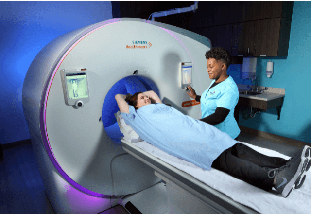
The primary goal of dental radiography in companion animals is early detection and accurate assessment of dental conditions. This helps in formulating a tailored treatment plan that minimizes pain and discomfort for the pet while ensuring effective management of oral health issues. The procedure typically involves positioning the animal’s head or mouth on an X-ray table, using a bite block to stabilize the jaws, and capturing images from multiple angles.
For quality assurance in dental radiography testing, it is essential to use equipment that meets international standards for precision and safety. This includes ensuring that the radiation dose delivered during imaging is kept as low as reasonably achievable (ALARA) to minimize potential risks to both the animal and the operator. Additionally, the images should be interpreted by a qualified veterinarian with expertise in radiology.
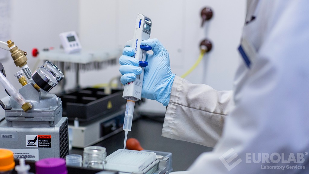
Before conducting dental radiography tests, proper specimen preparation is crucial. This involves ensuring that the patient is comfortable and stable during the procedure to obtain clear images. The use of appropriate contrast agents or dyes can enhance image clarity further. Post-processing techniques such as digital enhancement may also be employed to improve diagnostic accuracy.
When selecting a service provider for dental radiography testing, several factors should be considered:
- Experience and expertise of the veterinary team
- Adequate equipment calibration and maintenance
- Patient handling protocols ensuring minimal stress
- Use of advanced imaging technologies like cone beam computed tomography (CBCT)
- Compliance with relevant standards and guidelines
The application of dental radiography extends beyond just diagnosis; it also supports ongoing monitoring of treatment efficacy. Regular follow-up images can help track the progression or resolution of conditions over time, allowing for timely interventions if necessary.
Apart from routine check-ups, dental radiography may be recommended under specific circumstances:
- When there are unexplained symptoms like pain, swelling, or discharge
- To evaluate suspected fractures or luxations in the jaw bones
- In cases of chronic periodontitis where deep pockets or abscesses need to be assessed
- As part of comprehensive geriatric evaluations for older pets
In conclusion, dental radiography testing is an indispensable tool in modern veterinary practice. By providing detailed insights into the internal structures of companion animals' mouths, this service helps ensure optimal care and well-being.
| Applied Standards | Description |
|---|---|
| ISO 17634 | Quality requirements for medical imaging equipment used in veterinary medicine |
| AAMI PB26 | Packaging and sterilization of medical devices used in animal hospitals |
| ASTM F2857 | Performance requirements for portable dental X-ray systems |
| IEC 60601-1 | General safety requirements for electrical equipment used in veterinary medicine |
Benefits
Dental radiography offers numerous advantages over visual inspections alone, making it an invaluable asset in the field of companion animal dentistry. One key benefit is its ability to reveal hidden pathologies that would otherwise go unnoticed during a routine examination. This early detection capability allows for more effective treatment planning and intervention.
Another significant advantage lies in its role as a diagnostic tool during complex surgical procedures involving the maxillofacial region. By providing precise anatomical information, dental radiography aids surgeons in making informed decisions regarding incisions, resections, or other interventions. This not only enhances procedural accuracy but also reduces the risk of complications.
From an ethical standpoint, using dental radiography ensures humane treatment by avoiding unnecessary exploratory surgeries that could otherwise be avoided through non-invasive imaging techniques. Moreover, it promotes better patient outcomes by enabling proactive management strategies based on accurate diagnostic information.
The long-term benefits extend to improved quality of life for companion animals by preventing chronic pain and discomfort associated with untreated dental diseases. Regular monitoring via radiography contributes significantly towards maintaining overall health and longevity.


