Digital Subtraction Angiography in Veterinary Surgery
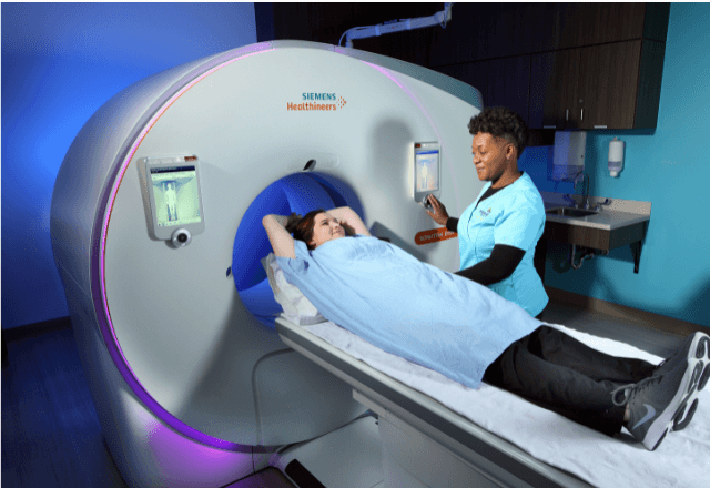
Digital Subtraction Angiography (DSA) is a sophisticated imaging technique widely used across human medicine but also increasingly applied in veterinary surgery. This advanced diagnostic method helps visualize blood vessels and their anomalies, which is crucial for accurate diagnosis and effective treatment planning. In the context of veterinary surgery, DSA provides detailed images that can guide surgeons during complex procedures involving arteries or veins.
The procedure involves injecting a contrast agent into the blood vessel under examination and capturing multiple X-ray images. These images are then processed by subtracting the background image to highlight only the vessels containing the contrast material. This process allows for precise identification of abnormalities such as blockages, aneurysms, or abnormal vascular structures.
DSA is particularly useful in veterinary applications where detailed visualization of blood flow and vessel architecture can significantly improve surgical outcomes. It enables surgeons to plan procedures with greater accuracy, minimizing risks associated with invasive interventions. The technology supports a wide range of veterinary specialties including orthopedics, cardiology, neurology, and urology, among others.
The use of DSA in veterinary medicine is supported by stringent international standards such as ISO 5210-3:2016 which outlines the basic principles for radiological imaging. Compliance with these standards ensures that images are of high quality and consistency, providing reliable diagnostic information essential for accurate interpretation.
One key advantage of DSA in veterinary surgery is its ability to provide real-time visualization of blood flow dynamics. This capability allows for dynamic assessment during interventions, enhancing the precision of therapeutic actions. Additionally, the use of advanced imaging techniques like DSA helps reduce the need for exploratory surgeries by providing accurate preoperative assessments.
Another important aspect of DSA is its role in post-operative monitoring. By allowing repeated imaging over time, it aids in assessing healing progress and identifying any complications early on. This proactive approach can lead to better patient outcomes and reduced hospital stays.
The application of DSA extends beyond just diagnostic purposes; it also plays a vital part in training veterinary surgeons. Detailed images captured during procedures serve as valuable educational tools, helping new professionals refine their skills. Moreover, the availability of high-resolution images facilitates collaboration between different clinics or specialists who may be involved in complex cases.
While DSA offers numerous benefits, it is important to consider that it comes with certain limitations and risks. The procedure involves exposure to ionizing radiation which can pose health hazards if not managed properly. Therefore, strict adherence to safety protocols is crucial when performing this test. Additionally, the cost associated with acquiring and operating sophisticated imaging equipment might be a barrier for some practices.
In conclusion, Digital Subtraction Angiography represents an indispensable tool in modern veterinary surgery. Its ability to provide clear images of blood vessels makes it invaluable for both diagnostic purposes and surgical guidance. However, careful consideration must always be given to patient safety and cost factors when deciding whether DSA is appropriate for a particular case.
Benefits
- Precise visualization of blood vessels and their anomalies,
- Real-time assessment during interventions enhancing the precision of therapeutic actions,
- Detailed preoperative assessments reducing the need for exploratory surgeries,
- Useful post-operative monitoring aiding in early identification of complications,
- Valuable tools for training veterinary surgeons providing detailed images as educational aids,
- Compliance with international standards ensuring quality and consistency.
Why Choose This Test
The choice of Digital Subtraction Angiography in veterinary surgery is driven by its ability to provide unparalleled clarity regarding the state of blood vessels. This level of detail allows for more accurate diagnosis and treatment planning, thereby improving surgical outcomes significantly.
By opting for DSA, veterinarians can ensure that they have all necessary information at hand before proceeding with any surgical intervention. This comprehensive knowledge helps in making informed decisions about which approach would be most beneficial for the animal's condition. Furthermore, the real-time capabilities of DSA make it especially advantageous during complex procedures where immediate adjustments might be required based on what is seen during the procedure.
For those considering this option, another compelling reason lies in its role as an educational resource. High-resolution images captured during DSA can serve as excellent teaching materials for both novice and experienced veterinarians alike. Such resources foster continuous learning and improvement within the veterinary community, ultimately benefiting all animals under care.
The commitment to using advanced technologies like DSA aligns with broader trends towards more precise and less invasive treatments in veterinary medicine. By embracing such tools, practices demonstrate their dedication to providing state-of-the-art care while maintaining high standards of ethics and professionalism.
Competitive Advantage and Market Impact
The adoption of Digital Subtraction Angiography (DSA) by veterinary practices offers significant competitive advantages. In a market where competition is fierce, being able to offer this advanced diagnostic tool sets apart a practice from its peers. Clients trust providers who can deliver cutting-edge services, knowing that their pets will receive the best possible care.
From a strategic standpoint, integrating DSA into routine operations enhances the overall reputation of a veterinary clinic or hospital. It signals commitment to excellence and innovation, attracting both new clients seeking these specialized services and existing ones looking for comprehensive healthcare solutions. Such recognition can lead to increased market share and customer loyalty.
In terms of operational efficiency, incorporating DSA improves workflow by streamlining diagnostic processes. The precise information provided enables quicker diagnosis, reducing unnecessary exploratory surgeries and subsequent recovery times. This not only benefits the animals but also reduces costs for both the practice and owners.
The impact on patient care is profound as well. By allowing earlier identification of issues through detailed preoperative assessments, DSA contributes to better treatment outcomes. Animals experience less stress during procedures due to more accurate planning, leading to faster recoveries and improved quality of life post-surgery.
Moreover, the continuous learning opportunities provided by high-resolution images captured during DSA further enhance the expertise level within veterinary teams. This ongoing professional development ensures that practices remain at the forefront of their field, continually striving for improvements in patient care standards.
In summary, embracing Digital Subtraction Angiography not only strengthens a practice's competitive position but also positively influences its market presence by demonstrating commitment to innovation and excellence. It enhances operational efficiency while significantly improving patient care through advanced diagnostics and better treatment outcomes.


