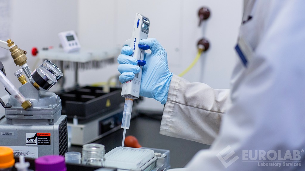
Immunohistochemistry Testing for Immune Cell Markers in Animals
The technique of immunohistochemistry (IHC) is a powerful tool utilized to visualize and characterize specific proteins, peptides, or antigens within tissues. In the context of clinical and healthcare testing, particularly in allergy and immunology, IHC has become indispensable for identifying immune cell markers in various animal species. This service involves the use of antibodies labeled with fluorescent or chromogenic substrates that bind specifically to target antigens present on the surface of cells or within tissues.
This methodology allows researchers and clinicians to not only visualize but also quantify the presence and distribution of specific immune cell types such as T-cells, B-cells, macrophages, dendritic cells, mast cells, and basophils. The process begins with tissue sampling from the animal under investigation. Typically, small biopsies are taken from relevant tissues or organs suspected to have an inflammatory or immunological response.
The sampled tissue is then fixed using formalin, embedded in paraffin wax, sectioned into thin slices (around 4-6 microns thick), and mounted on microscopic slides. These sections are subsequently deparaffinized, rehydrated, and subjected to antigen retrieval methods that enhance the visibility of antigens for antibody binding. Following this, a series of blocking steps is performed to prevent non-specific interactions between antibodies and tissue components.
The heart of the IHC process involves the application of primary antibodies directed against specific immune cell markers. These antibodies are typically conjugated with secondary antibodies labeled with fluorochromes or enzymes for visualization under fluorescence or light microscopy, respectively. Positive staining patterns indicate the presence of the targeted antigenic determinant on the cell surface or within the cytoplasm or nucleus.
The accuracy and reproducibility of IHC results depend heavily on various factors including antibody specificity, optimal antigen retrieval conditions, correct dilution ratios, appropriate control samples, and stringent quality assurance measures. Ensuring these parameters is crucial for generating reliable data that can contribute to a deeper understanding of immune responses in animal models.
One critical aspect of this testing method lies in its ability to correlate morphological changes observed within tissues with functional outcomes measured through physiological or biochemical assays. For instance, identifying increased numbers of T-cells infiltrating the lung tissue could suggest an allergic reaction, while elevated levels of interleukin-4 (IL-4) might further confirm Th2-mediated inflammation.
Another significant advantage of IHC is its capacity to differentiate between various subtypes of immune cells based on their unique surface markers. This capability enables more precise diagnoses and better treatment strategies tailored specifically for each condition. By providing detailed information about the nature, location, and extent of an immunological response, this technique serves as a cornerstone in translational research aimed at bridging laboratory findings with clinical applications.
For example, studies conducted on allergic asthma models have demonstrated how IHC can reveal enhanced infiltration of eosinophils into airway tissues. Such insights help refine therapeutic approaches targeting these specific cell populations. Similarly, understanding the role of regulatory T-cells in suppressing excessive inflammation can lead to novel immunosuppressive therapies designed to modulate aberrant immune responses.
In summary, immunohistochemistry testing for immune cell markers in animals plays a pivotal role in advancing our knowledge of allergic and immunological disorders across different species. Its versatility makes it an essential tool for researchers working on diverse projects ranging from basic science investigations into the mechanisms underlying disease processes to developing personalized medicine solutions aimed at improving patient outcomes.
Scope and Methodology
The scope of our immunohistochemistry testing encompasses a wide range of applications within allergy and immunology. We utilize this technique primarily to identify and quantify various immune cell markers in animal tissues, which is particularly useful for studying allergic responses, autoimmune diseases, and infectious conditions.
To ensure comprehensive coverage, we employ multiple staining techniques including indirect IHC, direct IHC, and double labeling methods. Each approach serves distinct purposes depending on the specific antigens being targeted. For instance, indirect IHC allows for simultaneous detection of several antigens by using biotinylated secondary antibodies, whereas direct IHC provides higher sensitivity due to its monospecific nature.
The methodology involves several key steps starting with tissue sampling followed by processing and embedding into paraffin blocks. The embedded tissues are then sliced into thin sections which undergo antigen retrieval processes tailored according to the type of antigen being analyzed. Afterward, primary antibodies specific for desired immune cell markers are applied, leading to subsequent incubation with secondary antibodies conjugated with fluorochromes or enzymes.
Following extensive washing steps designed to eliminate unbound reagents, sections are either counterstained using hematoxylin to highlight nuclei or mounted onto glass slides. Finally, images are captured under fluorescence or brightfield microscopes equipped with appropriate filters for optimal visualization of labeled antigens. These images form the basis from which detailed analyses regarding cell density and distribution patterns can be conducted.
Our laboratory adheres strictly to recognized international standards such as ISO 17025 and Good Laboratory Practices (GLP) guidelines ensuring accuracy, precision, and reliability of our results. Compliance with these stringent requirements guarantees that all tests performed meet the highest quality assurance criteria necessary for robust scientific inquiry.
Benefits
The benefits associated with immunohistochemistry testing for immune cell markers in animals extend far beyond mere identification; they offer profound advantages across multiple dimensions including research, development, and translational medicine. Firstly, this technique provides valuable insights into the dynamics of immune responses at both local and systemic levels within different species.
By enabling precise quantification and localization of specific immune cells, IHC facilitates the characterization of complex interactions between various cell populations involved in pathological processes. This capability enhances our understanding of disease mechanisms, paving the way for more targeted therapeutic interventions. For instance, elucidating the roles played by mast cells or basophils in allergic reactions can inform the design of anti-histaminic drugs.
Furthermore, IHC enables the study of immune cell trafficking patterns which are crucial for evaluating efficacy of novel treatments aimed at modulating inflammatory responses. By tracking the movement and accumulation of therapeutic agents within affected tissues, researchers gain critical information about their biodistribution characteristics.
The ability to differentiate between different subtypes of immune cells based on their unique surface markers also represents a major benefit. This precision allows for more accurate diagnosis of diseases characterized by aberrant immune responses such as autoimmune syndromes or cancers where specific cell populations become overactive or underrepresented.
From a practical perspective, IHC testing streamlines workflows significantly by reducing the need for multiple specialized analyses typically required when using other methods like flow cytometry. It achieves this through its integrated approach combining morphological assessment with biochemical profiling into single procedures performed on standardized tissue samples.
The cost-effectiveness of our services further enhances their utility making them accessible to a broader spectrum of investigators ranging from academic institutions conducting basic research to pharmaceutical companies engaged in drug discovery efforts.
Competitive Advantage and Market Impact
In an increasingly competitive landscape, our immunohistochemistry testing service stands out for several reasons that contribute significantly to its market position. One key advantage lies in the advanced technology used to perform these tests accurately and efficiently. Our state-of-the-art equipment allows us to offer services with high resolution images captured under fluorescence or brightfield microscopes equipped with appropriate filters.
Another significant factor is our commitment to adhering strictly to recognized international standards such as ISO 17025 and Good Laboratory Practices (GLP). This ensures that all tests performed meet the highest quality assurance criteria necessary for robust scientific inquiry. Compliance with these stringent requirements not only enhances reliability but also builds trust among clients seeking reliable results.
Our service is particularly valuable in areas where traditional methods might be less effective or impractical due to logistical challenges associated with handling live animals. For example, studying immune responses during acute infections or chronic inflammatory diseases requires careful sampling and processing of tissues from affected organs without causing additional stress to the animal subjects. Our expertise in this area ensures that such samples are handled delicately and analyzed accurately.
The versatility of our service also contributes to its competitive edge by catering to diverse needs across various sectors including academia, biotechnology, pharmaceuticals, and veterinary medicine. Whether it's elucidating mechanisms underlying allergic reactions or validating new biomarkers for diagnosis and monitoring purposes, we provide comprehensive solutions tailored specifically according to each client's requirements.
Moreover, our rigorous quality control measures ensure consistent performance across all tests conducted. This consistency is essential in maintaining credibility within the scientific community where reproducibility of results forms a cornerstone of valid research findings.
The impact on the market extends beyond mere service delivery; it lies in fostering innovation by enabling researchers to explore new avenues for understanding and treating immune-related disorders more effectively than ever before. By providing reliable data supported by robust methodologies, we contribute significantly towards advancing knowledge in allergy and immunology fields.


