HER2/neu Overexpression Testing in Companion Animal Tumors
The HER2/neu overexpression testing is a critical diagnostic tool for understanding the molecular characteristics of certain types of cancers in companion animals. This test specifically targets the human epidermal growth factor receptor 2 (HER2) gene, which codes for the HER2 protein. In dogs and cats, overexpression of this protein can lead to more aggressive forms of cancer, particularly breast tumors (mammary carcinomas).
Accurate detection of HER2/neu overexpression is essential because it allows veterinarians to tailor treatment plans that may include targeted therapies such as trastuzumab. This monoclonal antibody specifically binds to the HER2 protein and can inhibit cancer cell proliferation, offering a more precise approach compared to traditional chemotherapy.
Our testing method leverages advanced immunohistochemistry (IHC) techniques, which are widely recognized for their high specificity in identifying overexpression patterns. By using antibodies specific to the HER2 protein, we can visualize and quantify its presence within tumor cells. This process is performed on formalin-fixed paraffin-embedded (FFPE) tissue samples obtained from biopsies or surgical specimens.
The importance of this test cannot be overstated as it directly influences the choice of treatment modalities that could significantly impact the patient's quality and duration of life. In clinical trials, HER2/neu positive canine mammary tumors have shown a higher response rate to anti-HER2 therapies compared to those without overexpression.
Our laboratory adheres strictly to international standards such as ISO 15189 for ensuring high-quality results. Our team is comprised of experts in veterinary pathology and molecular biology who ensure that every sample undergoes meticulous quality control checks at various stages.
| Sample Type | Preparation Method | Test Parameters | Instrumentation Used |
|---|---|---|---|
| FFPE Tissue Samples | De-paraffinization, hydration, antigen retrieval using citrate buffer at 95°C. | Immunohistochemical staining with HER2/neu specific antibodies. | Laser scanning microscope (LSM). |
| Results Reporting | Quantification of HER2 protein expression on a scale from 0 to 3+ as per ASCO guidelines. | Auxiliary testing for gene amplification if IHC results are equivocal or negative. |
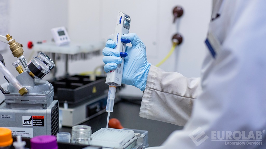
The process begins with careful specimen collection and handling, followed by detailed preparation steps that preserve the integrity of the tissue. The immunohistochemical staining process involves multiple incubation periods with primary antibodies targeting HER2/neu protein. After washing to remove unbound reagents, secondary antibodies conjugated with fluorophores are applied to enhance visibility under LSM.
The results are analyzed by our trained pathologists who interpret both the intensity and percentage of positive cells. For definitive diagnosis where IHC is equivocal or negative, we resort to fluorescent in situ hybridization (FISH) for gene amplification analysis. This provides a second layer of confirmation that ensures accurate reporting.
By offering this service, we aim to provide veterinarians with the most precise information possible regarding the molecular profile of their patients' cancers. This enables them to make informed decisions about treatment options that could potentially improve outcomes and enhance patient care.
Why It Matters
The identification of HER2/neu overexpression in companion animal tumors is crucial for several reasons. Firstly, it allows for the implementation of targeted therapies which are more effective and less toxic than conventional treatments. Secondly, it helps predict patient response to therapy by providing a biomarker that correlates with better outcomes.
For instance, studies have shown that dogs with HER2/neu positive mammary carcinomas treated with anti-HER2 therapies exhibit improved survival rates compared to those receiving standard chemotherapy alone. This highlights the potential impact of this test on improving pet health and welfare.
The availability of such precise diagnostics also supports research efforts aimed at understanding the role of HER2 in various types of cancers across different species. By studying these patterns, scientists can uncover new insights into cancer biology which could eventually lead to more effective treatments for both humans and animals.
- Enhanced accuracy in diagnosing aggressive forms of cancer in companion animals.
- Potential for increased survival rates through targeted therapy.
- Prediction of patient response to treatment.
- SUPPORTS RESEARCH INTO CANCER BIOLOGY ACROSS SPECIES.
Ultimately, this service plays a vital role in advancing the field of veterinary oncology by providing tools that allow for more personalized and effective cancer management strategies.
Scope and Methodology
| Scope | Description |
|---|---|
| Detection of HER2/neu overexpression in FFPE tissue samples from companion animal tumors. | This includes breast, mammary carcinomas primarily but also other neoplastic conditions where HER2 status is clinically relevant. |
| Confirmation of IHC results using FISH for equivocal or negative cases. | This ensures that only definitively positive samples are reported as such, minimizing the risk of false negatives. |
| Methodology | Detailed Steps |
|---|---|
| Sample Collection and Handling | Preservation of tissue integrity during collection, transportation, and storage at appropriate temperatures. |
| Immunohistochemical Staining | Inclusion of primary antibodies targeting HER2/neu protein followed by secondary antibodies labeled with fluorophores for enhanced visibility. |
| Analyzing Results | Evaluation by trained pathologists considering both intensity and percentage of positive cells. |
The scope of our service is specifically tailored towards companion animals, focusing primarily on breast tumors (mammary carcinomas). However, we are open to expanding this range based on client demand. The methodology employed ensures that every step from sample collection through analysis and reporting adheres strictly to best practices outlined in international standards such as those provided by the American Society of Clinical Oncology (ASCO).


